Petri Dishes with Nanotopography structure for cell culture
Recent results of Stem Cells studies claims that nanotopography could influence cell behavior – growth and differentiation.
Nanotopography Petri dishes are standard cell culture dishes, modified to have nanoscale features on the bottom. Nanotopography is embedded directly to the surface of dish with no additional layers.

Base Dish: SPL 10060
Material: Crystal grade Polystyrene
Dish Size: 60 mm X 15 mm
Total Area: 21.5 cm2
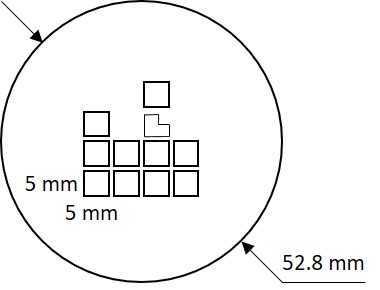
Number of Different Nanotopographies: 11
Nanotopography Area: 0.25 cm2 each square
Nanotopography Types: holes and lines
Typical sizes: 200~1100 nm

Preliminary studies of adipose-derived stem cells on a 5 different nanotopographies within one Nano Petri dish demonstrate difference between cell fate depending on the type of nanostructure.
